

OPTICAL COHERENCE TOMOGRAPHY
Optical Coherence Tomography (OCT) is a relatively new imaging technology that acquires high resolution images of the internal structures of the eye.
OCT uses light to image ocular tissue in a cross-sectional view. It has a resolution of 2-3 microns; 100 times finer than traditional ocular imaging techniques such as ultrasound, MRI, and CT.
The images acquired are a useful tool for assessing ocular diseases of the macula, optic nerve, and anterior structures of the eye, and for monitoring response to treatment.
The OCT scan is a brief, non-invasive imaging test. It typically takes less than 5 minutes to obtain the scans, and there are no associated risks. Contact Mohawk Valley Retina today for an eye disease diagnosis.
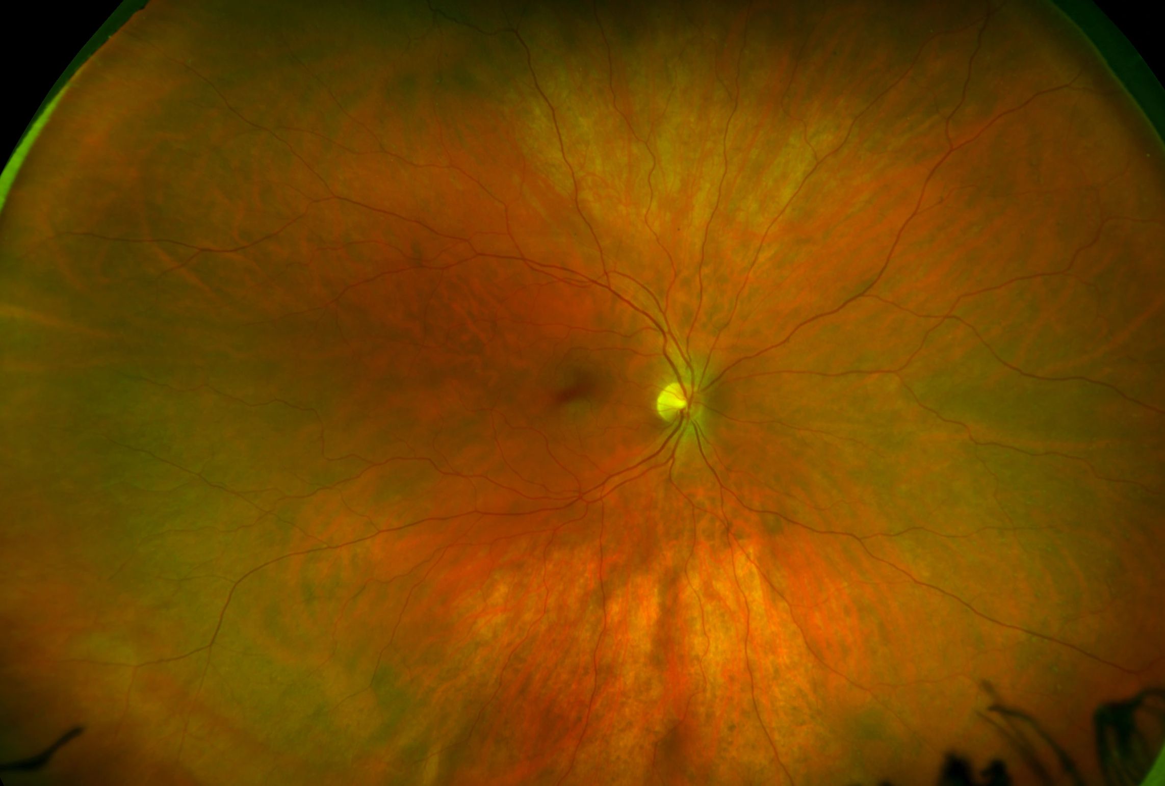
WIDE FIELD FUNDUS PHOTOGRAPHY
Fundus Photography involves capturing photographs of the back of the eye. Along with visualizing the macula, optic nerve, and retinal vessels, our wide field camera enables us to visualize the far peripheral retina.
.jpeg)
.jpeg)
.jpeg)
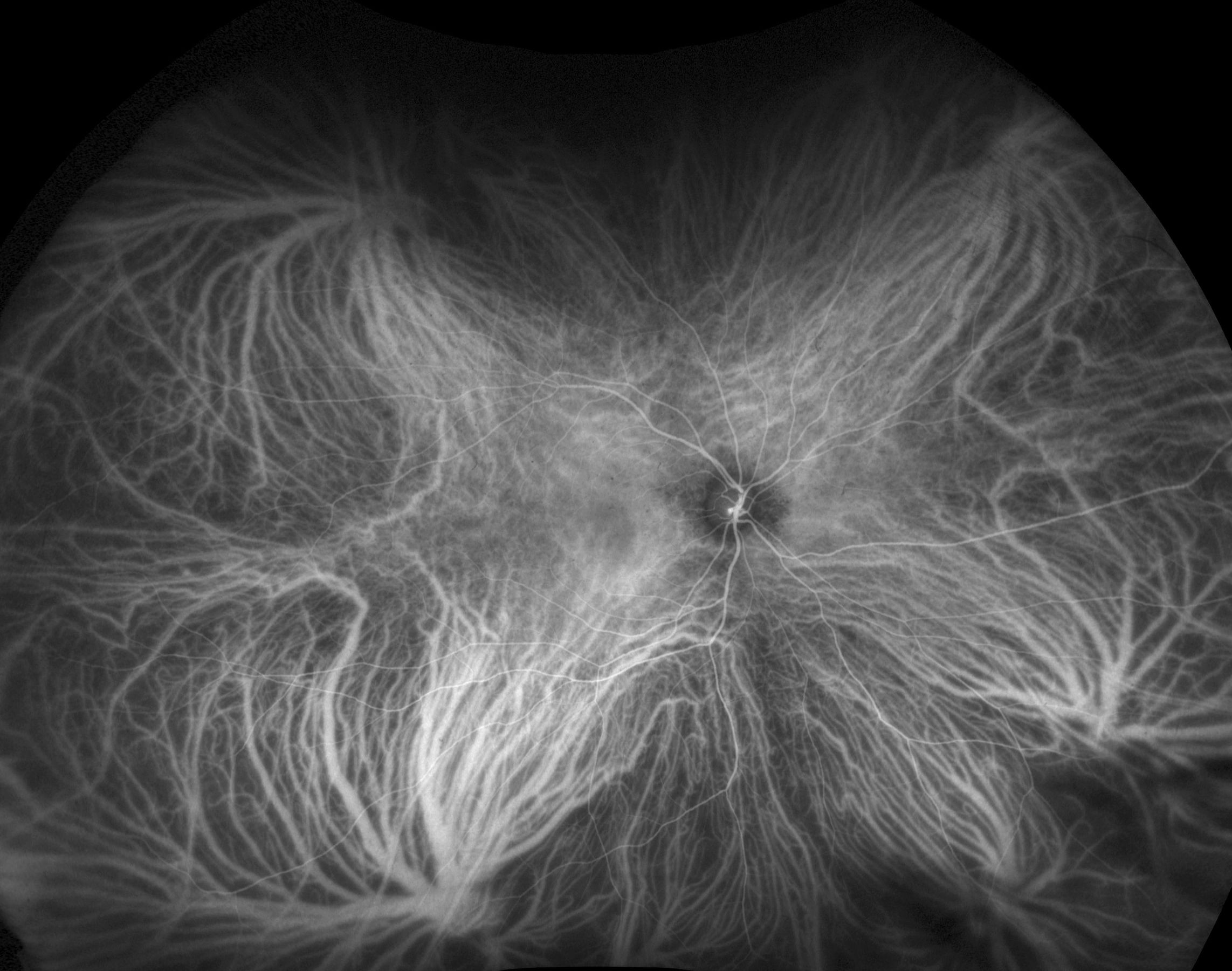
FLUORESCEIN ANGIOGRAPHY
Fluorescein Angiography is a photographic technique used for evaluating retinal circulation. During this test, a fluorescent medication is injected into a hand or arm vein. As the medication quickly travels to the eye, digital photographs are taken, which are then analyzed by the physician.
Vascular occlusion, wet macular degeneration, macular edema, diabetic retinopathy, and optic nerve disease all present with an abnormal fluorescent pattern on fluorescein angiography.
_1.jpeg)
INDOCYANINE GREEN ANGIOGRAPHY
Indocyanine Green Angiography (ICG) is a photographic technique used for evaluating circulation of the choroid, the layer of blood vessels which lies beneath the retina. During this test, ICG dye is injected into a hand or arm vein. As the dye quickly travels to the eye, digital photographs are taken, which are then analyzed by the physician.
_2.jpeg)
B-SCAN ULTRASONOGRAPHY
B-Scan Ultrasonography, or ophthalmic ultrasound, utilizes sound waves to image ocular tissue. High frequency sound waves travel through the eye and are reflected by ocular structures. These reflections form an image of the eye. Importantly, sound travels through structures that are opaque to light such as blood and dense cataract.
Ophthalmic ultrasound is a useful technique to image the posterior structures of the eye when the view is otherwise obscured by vitreous hemorrhage or cataract. Retinal tear or detachment, vitreous hemorrhage, and ocular tumors are visualized by ultrasonography.
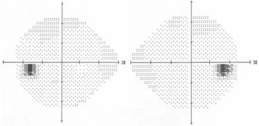
HUMPHREY VISUAL FIELD
Humphrey Visual Field (HVF) testing is a method of measuring not only central vision, but a patient's entire scope of vision. Importantly, HVF can detect vision loss that may be unnoticed otherwise. It is very useful in monitoring glaucoma patients and those with damage to the occipital cortex of the brain. HVF is also useful to assess the vision in macular disease and optic neuropathy.
During this test the patient looks at a central target. The automated perimetry test will briefly flash small lights on a white background. The patient presses a button whenever a light is seen. A computer assesses the entire field of vision and patterns of visual field loss can then be detected and measured. The test typically lasts 6-10 minutes per eye, and there are no associated risks.
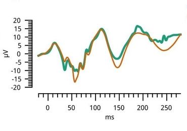
ELECTRORETINOGRAPHY
Electroretinography (ERG) measures the electrical responses of various cell types in the retina, including the photoreceptors (rods and cones), inner retinal cells (bipolar and amacrine cells), and the ganglion cells.
During the test, electrodes are placed on the skin beneath the eyes to measure the electrical responses to light as the patient's eyes are exposed to standardized stimuli.
VISUAL EVOKED POTENTIAL
Visual Evoked Potential (VEP) measures the electrical response of the brain's primary visual cortex to a visual stimulus. Three electrodes will be used to measure the electrical response. One electrode, which measures the response itself, goes over the primary visual cortex at the back of the head. Another electrode is placed at a reference location, typically on the forehead. The third electrode, for grounding, goes on the ear. Call (315) 732-0995 today for an eye disease diagnosis.
_1.jpeg)