
Central Retinal Vein Occlusion (CRVO) occurs when the main retinal vein is blocked. Because blood can no longer travel smoothly out of the retina, the retina fills with blood and becomes swollen. If blockage is partial, the retina may continue to function adequately. In more severe blockages, permanent retinal damage with loss of vision may occur. CRVO's are most common in people with high blood pressure, diabetes, or glaucoma. Occasionally, damage to the retina results in abnormal new vessel growth. Further visual loss may develop if the abnormal blood vessels break and bleed into the vitreous cavity, causing a vitreous hemorrhage.
Symptoms
Patients with CRVO usually experience blurred vision from retinal hemorrhage and swelling. Spots, strands, or curtains in the vision may occur due to this condition. Eye pain may be caused by neovascular glaucoma.Evaluation
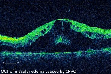
In addition to a dilated eye exam, a Fluorescein Angiogram may be required during which dye is injected into a vein in the arm. Digital images of the retina are obtained as the dye passes through the eye. Blocked or abnormal blood vessels then become detectable to the physician. Optical Coherence Tomography (OCT), a scanning laser which images a slice or cross section of the retina, may be performed to determine if macular edema is present. These tests aid in diagnosis to help establish the type of treatment needed.
Treatment
Intravitreal Drug Therapy improves the outcome in patients with CRVO. This procedure involves placing medication into the vitreous or main cavity of the eye. The injected medication acts upon damaged blood vessels to reduce leakage and resolve macular edema. Intravitreal drug therapy may also be recommended to cause regression of new blood vessels. The therapy may involve ongoing treatments, sometimes over many years.
Laser Treatment is often required when CRVO causes complications such as abnormal new blood vessel growth. Laser treatment is used to reverse growth of the new blood vessels. If untreated, these abnormal vessels can cause vitreous hemorrhage and glaucoma. Laser may be applied in several sessions.
_5.jpeg)
BRANCH RETINAL VEIN OCCLUSION
Branch Retinal Vein Occlusion (BRVO) is similar to a central retinal vein occlusion, but affecting only a portion of the retina. The condition occurs when a branch of the retinal vein becomes blocked, usually occurring where a retinal artery crosses over and compresses a retinal vein. The segment of retina drained by the blocked vein becomes swollen with blood. Where the vessel is blocked, leakage causing retinal swelling often occurs. Swelling in the central retina is called macular edema, which can result in loss of vision. As in CRVO, vision loss may also occur if abnormal blood vessels grow in the front or back of the eye.
Symptoms
Patients with BRVO often have blurred vision from retinal hemorrhage or macular edema. Spots, strands, or a curtain may occasionally appear due to vitreous hemorrhage. Eye pain may also be caused by neovascular glaucoma.Evaluation
In addition to the dilated eye exam, a Fluorescein Angiogram may be required. During this procedure, dye is injected into a vein in the arm. Digital images are then obtained of the retina as the dye passes through the eye, making the blocked or abnormal blood vessels detectable to the physician. Optical Coherence Tomography (OCT), a scanning laser that images a slice or cross section of the retina, may be performed to determine if macular edema is present. These tests aid in diagnosis, helping to indicate the need for treatment.
Treatment
Vision loss due to macular edema often requires treatment. There are two treatments available, which are sometimes used in combination:
Laser Treatment decreases the leakage that causes macular edema. Studies have shown that patients who receive laser for macular edema are twice as likely to regain good vision. If abnormal blood vessels are growing in the eye, more extensive laser treatment may be necessary.
Intravitreal Drug Therapy involves placing medication into the vitreous, or main cavity, of the eye. The injected medication acts upon damaged blood vessels to reduce leakage and resolve macular edema. Intravitreal injections are the most effective way of treating this condition and improving vision. Intravitreal drug therapy may involve ongoing treatments, sometimes for a number of years.